BLOOD THINNING MEDICATION’S PART 3: STATINS: WHAT ARE THEY AND HOW ARE THEY USED?

Over the past two weeks, I have described the blood thinning medications that vascular surgeons use to treat patients: Anticoagulant and Antiplatelet medications. This week, I want to spend time talking about another important medication that primary care physicians and vascular surgeons both agree helps to improve patient outcomes: Statins.

Statins refer to a general class of drugs that inhibit a special enzyme involved in the production of cholesterol within the body: 3-hydroxy-3-methylglutaryl-Coenzyme A, otherwise known as HMG-CoA.  HMG-CoA catalyzes the first reaction in a series of almost 30 individual reactions to create molecules of cholesterol!  As a vascular surgeon I sometimes prescribe Statins to my patients with high cholesterol to help my patients manage their cholesterol levels.

Cholesterol:

A high cholesterol level is one of the most common medical problems that patients I see have.  As a vascular surgeon it is important for me to understand when a patient has high cholesterol because of the following fact:

A high cholesterol level in the blood may result in a narrowing within the blood vessels of the heart, legs, and neck.  When these narrowing’s form they can cause heart attacks, ulcers and pain in the feet, and strokes, respectively.

In numerous clinical trials, the administration of statins has been shown to decrease the rate of death, heart attack, and stroke in patients at elevated risk of these medical problems due to high blood pressure, history of smoking, and diabetes.  The general group of statins includes medications like atorvastatin, simvastatin, and rosuvastatin.

At every office visit, I check my patients’ medical history to make sure that their cholesterol levels are being properly managed to ensure the best possible outcomes. Typically, when a patient presents in my office with a history of the following medical conditions, I will often prescribe statins as part of a larger approach to managing their cardiovascular disease:

  • History of heart attack, peripheral vascular disease, or stroke
  • History of diabetes mellitus over the age of 45

Vascular disease is both complex and difficult to manage. At South Bay Vascular Center and Vein Institute our physicians have been specially trained to understand “WHY” a problem exists so that our patients have real solutions to living a healthy life. Proper medical and surgical management of our patient involves a deep understanding of our patient’s medical history of which statins may play an important part. If you are currently on a statin or your referring physician believes a statin may be part of a larger approach to your circulatory health, please feel free to schedule an appointment with our office so that we can help you to evaluate this important decision with the context of your overall vascular health. Moreover, if you have a history of ulcers or pain in your feet or stroke, please do not hesitate to call my office today to schedule a consultation to determine if statins can be prescribed as part of your overall care and treatment!

Dr. Ignatius H. Lau: Vascular Surgeon

Dr. Ignatius Lau grew up in Portland, Oregon. He attended the University of Washington in Seattle for college and Stony Brook University in Long Island for medical school. He then went on to train in vascular surgery at Mount Sinai Hospital in New York City. During his time in New York, he performed over 1600 cases involving aortic, peripheral, venous, and carotid surgery. He has a special interest in limb salvage and treating patients with peripheral arterial disease and has extensive training and experience in treating the full spectrum of vascular diseases. Dr Lau was also very active in medical research during his training, ultimately finishing with twelve peer reviewed manuscripts. During his training in New York he met his wife, Lisa, who is a practicing endodontist. Together they love to hike, try new restaurants, and travel.

To see what our patients are saying about us, follow the link below to read our reviews.

https://www.google.com/search?q=south+bay+vasculr&rlz=1C1CHBD_enUS878US878&oq=&aqs=chrome.0.69i59i450l8.2997409j0j7&sourceid=chrome&ie=UTF-8#lrd=0x808e34eecfbc0653:0xb9aa2de7f50ba6a5,1,,,

South Bay Vascular Center and Vein Institute is Silicon Valley’s largest and most trusted Vascular Surgery practice.  Serving South Bay communities for over 26 years, Dr Kokinos and her Colleague, Dr Ignatius Lau are the region’s foremost experts in advanced vascular care and provide innovative care for patients suffering from circulation relation problems. At South Bay Vascular Center and Vein Institute our job is to understand the “Why” so that you have real solutions to living a healthy life. Call us today at 408-376-3626 or visit our website at www.southbayvascular.com to learn about what makes us the most referred to vascular surgery clinic in Silicon Valley.
BLOOD THINNING MEDICATIONS PART 2: ANTIPLATELETS: PLAVIX AND ASPIRIN. WHAT ARE THEY AND HOW ARE THEY USED?

Last week I began a description of the medications that vascular surgeons use with a blog post on drugs used for anticoagulation.  This week, I want to describe a group of medications that also thin the blood, albeit through a different mechanism.  As discussed last week, the coagulation cascade works to turn the blood from a liquid to a solid.  A special group of cells in the blood mixed with red blood cells called platelets works simultaneously with the coagulation cascade to serve as the glue so to speak between clumps of red blood cells.  This bond that platelets facilitate with clotted blood helps clot to attach and build upon other collections of clot, thereby facilitating the control of bleeding that the clotting system was designed for.

Obviously, in the case of vascular surgery we often times want to prevent blood clotting from occurring.  The main agents we use are aspirin and plavix (clopidogrel).  In the early 2000s literature from interventional cardiology for minimally invasive heart procedures found that placing patients on aspirin and plavix together reduced the incidence of recurrent heart attacks as well as death.  This literature from cardiology has been extrapolated to the lower extremity circulation and allows the interventions we perform in the lower extremities to fix blood flow to stay open for a long period of time.  In addition to the use of aspirin and plavix for lower extremity arterial blockages, we also use it in patients who have had a stroke in order to help prevent them from having another stroke.

Aspirin is a low strength blood thinner that patients can take orally as an 81 mg tablet once a day.  In the body aspirin blocks a specific enzyme called cyclooxygenase.  When cyclooxygenase is inhibited, the enzyme cannot help to produce chemicals in the blood called prostaglandins.  Prostaglandins are normally responsible for creating an environment that stimulates the clotting of blood via complex but mild mechanisms.  Studies have shown generally that patients with cardiovascular disease have a lower incidence of death, stroke, and heart attack over time than patients who do not take aspirin.  In general, I tend to make sure that all my patients who have peripheral vascular disease take aspirin as a general preventative measure.

Plavix is a much higher strength blood thinner that binds directly to platelets and completely inhibits their function.  When used in concert with aspirin, the blood becomes thin enough to prevent the recurrent blockage of vessels that we have opened up.

Overall the large majority of my patients who receive procedures to fix blood flow to the foot are placed on aspirin and plavix.  If you think you might benefit from these medications or are in need of a procedure to fix the blood flow to your feet or brain, please do not hesitate to call my office to schedule an appointment!


South Bay Vascular Center and Vein Institute is Silicon Valley’s largest and most trusted Vascular Surgery practice.  Serving South Bay communities for over 26 years, Dr Kokinos and her Colleague, Dr Ignatius Lau are the region’s foremost experts in advanced vascular care and provide innovative care for patients suffering from circulation relation problems. At South Bay Vascular Center and Vein Institute our job is to understand the “Why” so that you have real solutions to living a healthy life. Call us today at 408-376-3626 or visit our website at www.southbayvascular.com to learn about what makes us the most referred to vascular surgery clinic in Silicon Valley.
CLICK ON THE LINK BELOW TO READ HOW OUR PATIENTS DESCRIBE THEIR EXPERIENCE AT SOUTH BAY VASCULAR.

https://www.google.com/search?q=south+bay+vascular+center&rlz=1C1GGRV_enUS748US759&oq=&aqs=chrome.0.69i59i450.91302019j0j7&sourceid=chrome&ie=UTF-8#lrd=0x808e34eecfbc0653:0xb9aa2de7f50ba6a5,1,,,


PART 3: WHY DO MY TOES, ANKLES AND FEET HURT AT NIGHT? PERIPHERAL ARTERIAL DISEASE

In part one- and two of our four-part series on “Why do my feet hurt at night”, I discussed Venous disease and Raynaud’s Syndrome. In today’s discussion I want to focus on an even more common reason for why patients may suffer from leg pain at night: Peripheral arterial disease (otherwise known as PAD.)

As we’ve discussed in our earlier blogs, there are two kinds of blood vessels: Arteries and veins.

  • Arteries bring blood from the heart to the foot and toes under high pressure while
  • Veins bring the blood back from the foot to the heart under low pressure.

Over the course of anyone’s lifetime, blockages may build up in the arteries between the heart and the feet, especially in the pelvis and the legs.  Risk factors that increase the risk of formation of these blockages include:

  • Obesity
  • High blood pressure
  • High cholesterol
  • Diabetes
  • History of smoking tobacco.

As you may imagine, the tissue within the foot needs a certain amount of blood to supply the nutrients and oxygen necessary to keep the cells within the foot living.  Though a single, short segment blockage of the arteries in the leg may not result in any problems, multiple blockages from the pelvis to the foot may can make it difficult or even impossible for the cells within the foot to survive this lack of nutrients and oxygen.

What Does PAD Feel Like?

Typically, patients notice a gradual onset of pain in the forefoot and toes over the course of a few weeks to months.  As their blockages worsen, their pain becomes more severe.  Pain from PAD occurs in the forefoot and toes because those parts of the body are the furthest away from the heart and thus any blockage along the way from the heart to the toes can contribute to the lack of blood flow. One classic symptom of PAD pain is a burning, tingling, or numb sensation that can be partially relieved by hanging your foot off the bed or a chair.  In this position with the foot hanging down, gravity assists the flow of blood to the foot and toes and relieves the pain.  Typically, patients complain that when they lay flat at night, the pain in the forefoot and toes or just the foot in general will wake them up.  I always specifically ask patients if they are woken up at night by the pain.  If they are woken up by the pain, then I know the PAD must be very severe.

In some patients, the lack of blood flow is so diminished that there is also the development of ulcers, or defects in the skin with exposure of underlying fatty tissue, bone, and muscle or gangrene, the death of the tissue itself.

Even though PAD is a very severe and life limiting disease, specially trained vascular surgeons have many tools to combat it!  We can do a minimally invasive procedure called an angiogram that I described in an earlier blog post that you can access here!  Essentially, I access the diseased blood vessel with a needle and through that needle use contrast and x-ray to see where the blockages are.  Then, using balloons, stents, or self-expanding metal tubes, it is possible to re-open the blockages to re-establish blood flow to the impacted area.  For severe blockages, I also use a device called atherectomy that allows me to cut through and to remove the plaque responsible for blocking this vessel.  The procedure on average takes 1.5 to 2.5 hours and most patients can go home the same day.

Does foot pain wake you up at night? If you are experiencing pain in your foot or toes, please do not hesitate to contact my office at 408-376-3626 to schedule a clinic appointment today!

We Can Help!

Dr. Ignatius H. Lau

Vascular Surgeon

Dr. Ignatius Lau grew up in Portland, Oregon. He attended the University of Washington in Seattle for college and Stony Brook University in Long Island for medical school. He then went on to train in vascular surgery at Mount Sinai Hospital in New York City. During his time in New York, he performed over 1600 cases involving aortic, peripheral, venous, and carotid surgery. He has a special interest in limb salvage and treating patients with peripheral arterial disease and has extensive training and experience in treating the full spectrum of vascular diseases. Dr Lau was also very active in medical research during his training, ultimately finishing with twelve peer reviewed manuscripts. During his training in New York he met his wife, Lisa, who is a practicing endodontist. Together they love to hike, try new restaurants, and travel.

PART 2: WHY DO MY TOES, ANKLES, AND FEET HURT AT NIGHT? RAYNAUD’S SYNDROME

Last week I wrote the first part of a four-part series about foot pain at night.  Specifically, I talked about venous disease as a cause of foot pain.

Today, I want to talk about another common cause of foot pain at night: Raynaud’s Syndrome. 

As a quick review, last week we talked about the two main types of blood vessels in our bodies: arteries and veins.  Arteries bring blood from the heart to the feet under high pressure.  Veins bring blood back from the foot to the heart under low pressure.

Raynaud’s Syndrome is a disease of the arteries, particularly the small arteries, in the feet.

(Though Raynaud’s can also affect the hands as well). 

Before we dive into Raynaud’s Syndrome though, I need to briefly review another anatomical system in our bodies that is involved in Raynaud’s Syndrome: The nervous system.

In general, we have two broad types of nerves.

  1. There are nerves that are responsible for sensation and for moving muscle and
  2. There are nerves that are responsible for regulation of bodily function.

Diving a little bit deeper into the kinds of nerves that regulate bodily function, there are the

  • Sympathetic nervous system: The sympathetic nervous system, amongst other things, is responsible for opening blood vessels in the legs and arms and increasing blood flow when exercise is necessary
  • Parasympathetic nervous system: The parasympathetic nervous system in similar fashion opens the blood vessels leading to the intestines to allow for the digestion of food.

In patients with Raynaud’s Syndrome, sometimes their sympathetic nervous system does not function properly resulting in the constriction of blood vessels that lead to the hands, fingers, feet, and toes.  When it happens, blood vessels in the fingers and toes typically first turn blue because of the lack of oxygen; then white because of the lack of blood flow; and finally red, once the constriction ends and the blood flow returns to the fingers and toes.  During this constriction of the blood vessels, patients can have significant pain, numbness, tingling, and, in very severe cases, ulcers (defects in the skin with exposure of underlying fat and muscle) and gangrene (black and dead tissue).

Interestingly, environmental and social triggers play a significant role in Raynaud’s Syndrome episodes of vessel constriction.  Factors including stress, caffeine, alcohol or other drugs, and cold temperature can all induce a painful episode with lack of blood flow to the hands and feet.

Not much is known about the cause of Raynaud’s Syndrome or specifically why it happens.  Sometimes, Raynaud’s Syndrome can occur on its own without any other related disease.  In other cases, the patient has another inflammatory disorder like lupus or Sjogren’s Syndrome that is known to be highly associated with Raynaud’s Syndrome.

Though we do not fully understand the causes of Raynaud’s Syndrome there are many treatment strategies that can help.  Starting with behavioral modifications and certain blood pressure medications that can reduce spasm to specific procedures aimed to reduce the spasm of the vessels induced by the sympathetic nervous system, there is so much that I as a vascular surgeon can do to help patients with Raynaud’s Syndrome.  If you think that you might suffer from Raynaud’s Syndrome, please do not hesitate to call our office on 408-376-3626 to schedule an appointment. We Can Help!

Dr. Ignatius H. Lau

Vascular Surgeon

Dr. Ignatius Lau grew up in Portland, Oregon. He attended the University of Washington in Seattle for college and Stony Brook University in Long Island for medical school. He then went on to train in vascular surgery at Mount Sinai Hospital in New York City. During his time in New York, he performed over 1600 cases involving aortic, peripheral, venous, and carotid surgery. He has a special interest in limb salvage and treating patients with peripheral arterial disease and has extensive training and experience in treating the full spectrum of vascular diseases. Dr Lau was also very active in medical research during his training, ultimately finishing with twelve peer reviewed manuscripts. During his training in New York, he met his wife, Lisa, who is a practicing endodontist. Together they love to hike, try new restaurants, and travel.

FIXING BLOCKED ARTERIES

The most common disease that I treat as a vascular surgeon is a medical problem called peripheral vascular disease.  Specifically, peripheral refers to the legs, and vascular disease refers to blockages of the arteries.

Because peripheral vascular disease is the most common disease that I treat, I want to talk today about what I can do as a vascular surgeon to fix these kinds of blockages in the arteries!

First though, let me explain why blockages to the arteries of the legs are bad.  When not enough blood flow reaches the legs, patients can have pain in their calves or buttocks that limits their ability to walk longer than a city block.  When the blockages are very severe, they can also have pain in their feet and toes even when they are not moving or develop non-healing ulcers and gangrene.  When I open blockages in patients’ legs, I allow them to walk again, or I give them the ability to heal their ulcers and gangrene pain free.  The improvement in patient lives is the most exciting aspect of my job!

There are two main approaches to performing what we call revascularization. Revascularization refers to the opening of old passageways or creation of new passageways to bring blood directly from the heart to the foot without any obstruction.

  • Open Surgery: In the early decades of vascular surgery, namely the 1980s and 1990s the primary way to treat peripheral vascular disease was to perform an open surgical bypass.  If there was a blockage in the thigh, I would surgically expose above and below the blockage and take a synthetic tube or the patient’s own vein and then sew it above and below the blockage.  This would allow the flow of blood to bypass the blocked segment, hence, the reason we call this surgery a bypass!  Though this procedure does produce robust flow, it necessitates large surgical incisions that cause significant pain and are susceptible to poor wound healing and/or infection.


  • Endovascular Surgery: In the 2000s, an innovative approach called endovascular surgery started to become more widely used and is now actually standard of care today.  Indeed, I prefer to treat patients with peripheral vascular disease with endovascular surgery whenever possible.  We access the arterial system of the leg with a needle in the groin.  Through that needle we put in a strong wire and over that wire we can then advance several different instruments over catheters.  In general, there are three different techniques I can employ in my efforts to re-establish blood flow to an area that is no longer receiving blood due to some type of arterial blockage.
    • Balloons: The most basic instrument is a balloon that expands and breaks up the narrowing or blockage.
    • Stents: If the balloon does not work, we can use a self-expanding tube called a stent.
    • Atherectomy: If the stent does not work, we can use a special device called atherectomy.  Atherectomy involves the use of a device that can literally cut the plaque out of the artery and aspirate all the debris.


These three technologies allow the reopening of arteries from the toes to the pelvis and only necessitate a 2 mm puncture site in the skin of the groin to be used.  There is little to no risk of infection and the procedure can be performed several times over the course of the patient’s life.  On the other hand, surgical bypass can only be performed twice—three times—during the patient’s life due to the development of dense scar tissue after each operation.


If you have trouble walking because of pain in your buttocks or calves or have pain in your feet and toes or ulcers that will not heal, please do not hesitate to call my office today at 408-376-3626 to schedule an appointment.  The chances are that I will be able to help take your pain and ulcers away! We Can Help

www.southbayvascular.com

ANEURYSMS: WHAT ARE THEY AND WHY IS IT IMPORTANT TO KNOW IF YOU HAVE ONE?

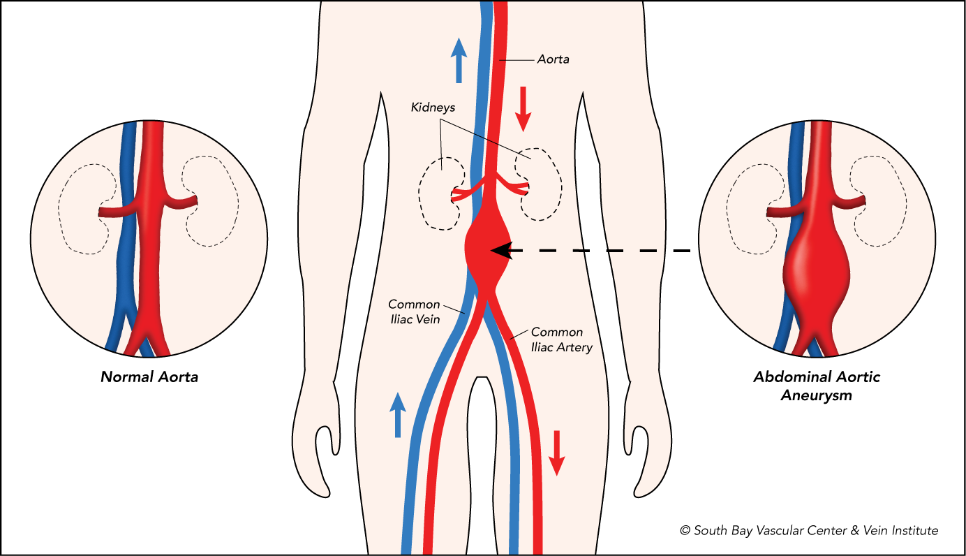
Of the many reasons that a patient can be referred to a vascular surgeon, one is that their referring physicians believes that their patient may have developed an aneurysm.  Aneurysms, typically speaking, form in arteries which are the specialized vessels that carry blood from the heart to the rest of the body under high pressure.  When the wall of an artery weakens, it can develop into a balloon-like dilation called an aneurysm. (See below). This most commonly occurs in the main blood vessel in your abdomen, which is called the abdominal aorta. When the blood vessels dilate to 50% greater than their normal diameter, the vessel has become an aneurysm.


Interestingly, the reason why aneurysms form is actually poorly understood.  We know that smoking cigarettes and a family history of aneurysms increases the risk that a patient will develop an aneurysm, but no direct cause-effect relationship has been established.  Diabetes mellitus, however, appears to have a protective effect.  That is, the presence of diabetes appears to help prevent aneurysms!  The other health risks of diabetes including neuropathy and infection, of course, make diabetes an undesirable health problem to have.

Aneurysms have two main catastrophic problems: Rupture and thromboembolization.  (to learn more about aneurysms visit our website at https://www.southbayvascular.com/aneurysms/  Rupture refers to a tear in the wall of the blood vessel resulting in a leakage of blood.  When aneurysms in the abdomen rupture, the amount of blood loss into abdomen can be life threatening and result in death within seconds.  Thromboembolization refers to the formation of blood clot within the aneurysm that then breaks off and goes somewhere else.  When aneurysm and associated blood clot form in the abdomen, these blood clots can break off and travel to the leg resulting in a lack of blood flow to the legs.  Such a sudden cessation of flow to the legs can result in the death of the foot and/or legs within hours.

As you can see arterial aneurysms can be very troublesome.  Luckily, vascular surgeons have many tools and instruments that we can use to treat these aneurysms in a minimally invasive way.  In my blog next week, I will go over the different options for treating aneurysms in the abdomen.  If you or someone you love have been told that you may have an aneurysm, please call our office immediately to schedule an appointment for a full vascular work-up. 408-376-3626

We Can Help!


WHAT IS “ENDOVASCULAR” SURGERY?

As a vascular surgeon, I perform two very different types of surgery: Open Surgery and Endovascular Surgery.

  • Open surgery is just as you might think!  It involves making an incision with a scalpel and using various tools like forceps, scissors, electrocautery, and needle drivers to expose and then repair specific structures.  Since the beginning of surgery itself, all surgeons, not just vascular surgeons, have been practicing open surgery.
  • Endovascular surgery is a recent development that only began in the 1990s.  Motivated by the desire to perform minimally invasive and less harmful interventions on the most sick and highest risk patients, pioneers in vascular surgery developed endovascular techniques.

Though the concept is simple, endovascular surgery now allows vascular surgeons to solve a multitude of problems with blood vessels very differently than they have in the past. Using advanced minimally invasive surgical techniques, vascular surgeons can treat many forms of vascular disease without the need to “cut-open” their patients to treat extraordinarily complex and in some cases life threatening diseases.

Endovascular surgical techniques utilize ultrasound (sound waves) to place the tip of a hollow needle into a blood vessel.  Once these hollow needles have been placed through the needle, we place a very stiff wire that allows us to advance large tubes called catheters into the vessel.  Catheters have many different functions depending on the situation and are used to re-establish blood flow in vessels that are diseased and or blocked.

For peripheral vascular disease, there are blockages in the legs of a patient that prevent blood flow from reaching the foot.  For peripheral vascular disease we can use balloons on the end of the catheter that are inflated to open a blockage.  If balloons do not work, then we can put a self-expanding metal mesh tube on the end of a catheter and then deploy it inside the blockage, thereby keeping it open.  There is also a technology called atherectomy which literally means cutting out plaque.  The atherectomy device has a cutting edge that sands down the plaque and then a suction function that removes all the debris.

For aneurysms, which are dilations of a blood vessel that can rupture, we use stent grafts, which are self-expanding metal mesh tubes that are lined with an impermeable fabric.  We place the stent graft above and below the aneurysm, thereby sealing it from the pressure created by the heart and preventing rupture.

At the end of these endovascular cases, the patient only has one or two 1 mm in size punctures over the arteries that were accessed.  Contrast this to the classic open surgeries where patients could have incisions up to 10 to 20 cm!  As a contemporary vascular surgeon, I take pride and joy in being able to offer both types of surgical interventions to my patients.  If you or your loved one may be suffering from vascular disease, please do not hesitate to call our office at 408-376-3626 to schedule an appointment today!

YOU HAVE A BLOOD CLOT. NOW WHAT?

One of the most common consultations that we as vascular surgeons receive is for deep vein thrombosis, otherwise known as blood clots.  Deep veins refer to the location of the vein that has the problem.  In the legs we have two main sets of veins: Deep and superficial.  Deep veins are within the muscle and close to the bones whereas superficial veins are close to the skin; hence their names!

Thrombosis refers to the process of the blood turning from a liquid into a solid form.  Normally, thrombosis occurs in response to an injury to stop bleeding from a wound.  However, when thrombosis occurs in the deep veins, it stops blood from being transported from the foot back to the heart.  The obstruction of flow results in swelling and pain below where the thrombus, or clot, has formed.

There are many causes of blood clots, but the most common causes include:

  • Traveling for extended periods of time in a sitting or resting position.
  • Trauma, including accidents which cause broken bones or bruises.
  • Narrowing of the veins of the pelvis, and
  • Specific genetic mutations that increase the likelihood of the blood forming clots. 
  • In older patients, another potential cause of blood clots without any other cause is cancer.  Cancer, unfortunately, can result in the release of chemicals and hormones within the body that make clotting more likely.

For patients who develop a blood clot in the context of traveling, narrowed pelvic veins, or having a traumatic injury to the affected leg, treatment involves a 3-month course of blood thinning medication followed by ultrasound tests to determine the stability of the clot.  If the symptoms improve and the clot remains stable, then the medication to thin the blood can be stopped at 3 months.

For travelers who are forced to sit for extended periods without being able to move, making an effort to walk around intermittently throughout the traveling can be helpful.  For example, on the plane, every 30 minutes stand up and walk up and down the aisle 3-4 times.  For patients who have a traumatic injury to their leg, the best choice would be to avoid, if possible, situations that led to the particular accident.  Finally, in patients with narrowed pelvic veins, I as a vascular surgeon can help relieve the narrowing with a special minimally invasive procedure called a venogram.  In the procedure, I place a self-expanding tube called a stent into the compressed vein via a small 2 mm puncture site in the groin to make sure the compressed vein remains open.

For those patients who did not have a traumatic accident, narrowed pelvic veins, recent travel, or a high suspicion of cancer, referral to a hematologist, or a doctor who specializes in disorders of the blood, can be helpful.  The hematologist can help order special genetic tests to determine if there is a genetic predisposition to forming clots.  In patients who do end up having genetic mutations, long-term placement on blood thinning medications can help prevent further events.

If you are anyone that you know is suffering from a blood clot, please do not hesitate to call our office on 408-376-3626 to schedule an appointment!  The expertise that vascular surgeons have in this area of medicine is critical to ensure a good outcome!

TRANSCAROTID ARTERY REVASCULARIZATION: THE FUTURE OF CAROTID SURGERY

One of the most gratifying problems we help treat for patients is carotid artery disease.  The carotid arteries supply the brain with blood.  In patients with high blood pressure, high cholesterol, diabetes, and a history of smoking, the carotid arteries can become narrowed and form blood clots that then go into the brain and cause strokes.  Traditionally, vascular surgeons have fixed this carotid artery narrowing’s with a surgery called a carotid endarterectomy.  The carotid artery is dissected out and cut open.  The plaque is then removed, and the carotid artery is sewn back together.

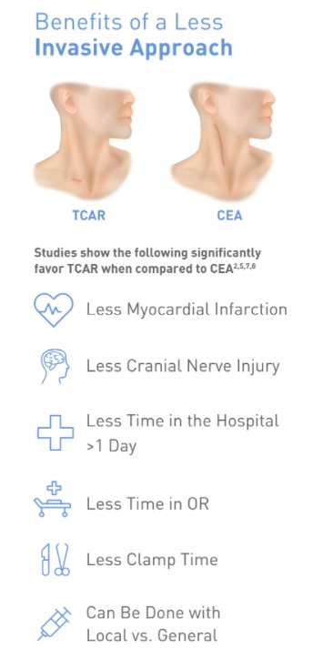
Over the past five years, however, I have had the opportunity to perform a new technique involving carotid stenting called trans carotid artery revascularization or TCAR.  Stents are self-expanding metal tubes that we can use in the carotid artery to stop clots from forming in narrowed areas.  TCAR uses a suction machine to reverse flow in the carotid artery during the placement of a stent to minimize the risk of inadvertent stroke during the procedure itself.  After having performed over 20 of these procedures in the past two years, I can say with confidence that the flow reversal and stenting that TCAR provides is a highly safe and successful way to treat carotid artery disease.  Furthermore, the length of the incision, postoperative pain, and risk of nerve damage and bleeding are all much less with TCAR than with CEA.

I am so happy to be able to offer this revolutionary, safe, and effective therapy to all my patients at South Bay Vascular Center.  Should you or anyone you know have any problems with your carotid arteries or a stroke, please do not hesitate to call us today at 408-376-3626 to schedule a consultation.

https://youtu.be/O32nDoovMPY]

WHY DON’T THE WOUNDS ON MY LEGS AND FEET HEAL ANYMORE?

Hi! Dr. Lau here again with South Bay Vascular. Today, I wanted to shed some light on one of the most common issues that we see in vascular surgery, non-healing wounds of the feet and toes. My hope is that after reading this blog you will have a better understanding of what causes leg wounds and ulcers; why sometimes they won’t heal on their own, and what we in vascular surgery can do to help them heal.

Ulcers, generally speaking, develop because of TWO main causes: nerve damage induced by diabetes and lack of blood flow. When patients have elevated levels of sugar in their blood with diabetes, the sugar forms toxic compounds that damage nerves in the foot. Consequently, the patient cannot feel injuries that would otherwise cause a normal patient to adjust position to stop the injury. With this loss of sensation ulcers form at the point of repeated injury that the patient cannot feel.

The second cause, a lack of blood flow, develops over decades in patients who smoke and who have high blood pressure and high cholesterol. These medical problems cause hardening and narrowing of the blood vessels, otherwise known as atherosclerosis.

In patients with diabetes, I coordinate carefully with my podiatrist colleagues who are experts in specialized footwear and surgical interventions that alleviate common areas of trauma. In patients with poor blood flow, I, as a vascular surgeon, can perform minimally invasive surgery to restore blood flow to the affected area. We use wires and specialized tubes to gain access to the blood vessels and then use balloons to break open the narrowing’s and self-expanding tubes called stents to keep the vessels open. Afterwards, we use a specialized x-ray machine called fluoroscopy to see that the narrowing’s have opened again to allow blood to flow back to the area. Patients usually leave to go home the same day with only a 2-millimeter puncture in their groin and are back to normal activity the day after surgery!

If you or anyone you know has a wound on their leg, foot or toe that hasn’t healed in more than two weeks, please call our office at 408 376 3626 to schedule an appointment.